Device-agnostic interface
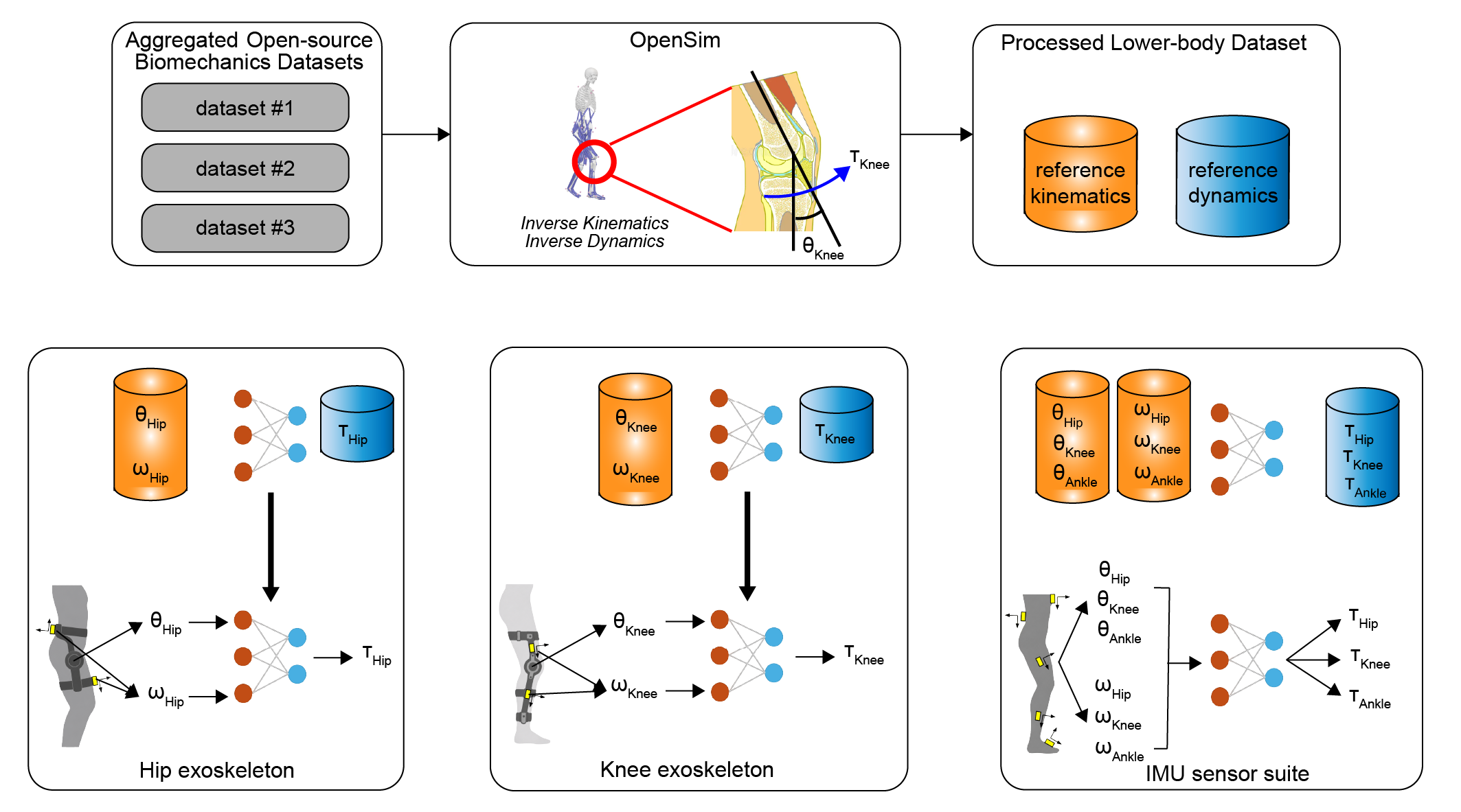
Joint angles and angular velocities form a shared biomechanical input domain, so each wearable only needs to produce compatible kinematics rather than matching another device’s raw sensors.
Joint angles and angular velocities serve as a shared intermediate representation, decoupling wearable sensing hardware from downstream biological joint moment estimation.
1Carnegie Mellon University, Mechanical Engineering
Joint angles and angular velocities form a shared biomechanical input domain, so each wearable only needs to produce compatible kinematics rather than matching another device’s raw sensors.
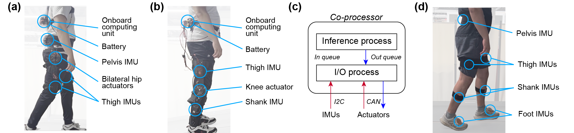
Causal TCN estimators are trained exclusively on public biomechanics datasets and applied without target-device training or fine-tuning to hip exo, knee exo, and IMU kinematics.
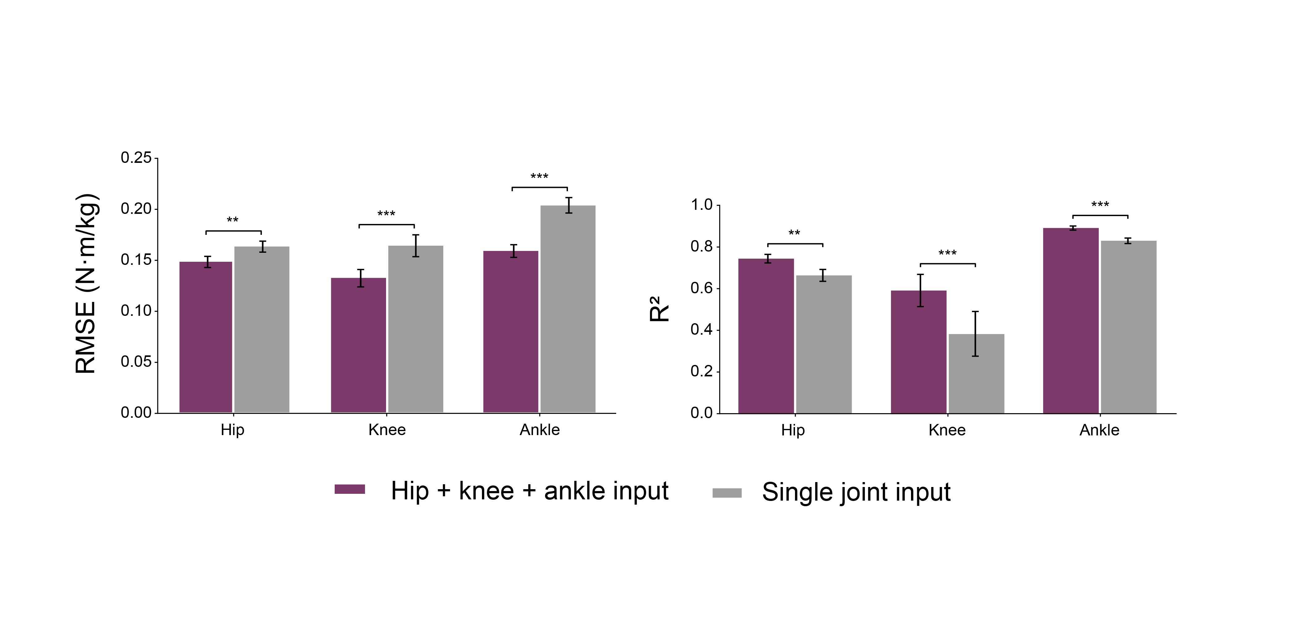
On the Awinda IMU suite, multi-joint (3-DoF) inputs cut mean RMSE by 17.2% versus single-joint models, with significant gains at hip, knee, and ankle.
Each wearable converts its sensors into joint kinematics; a shared estimator maps those kinematics to biological joint moments—trained once on open-source data, deployed across platforms.
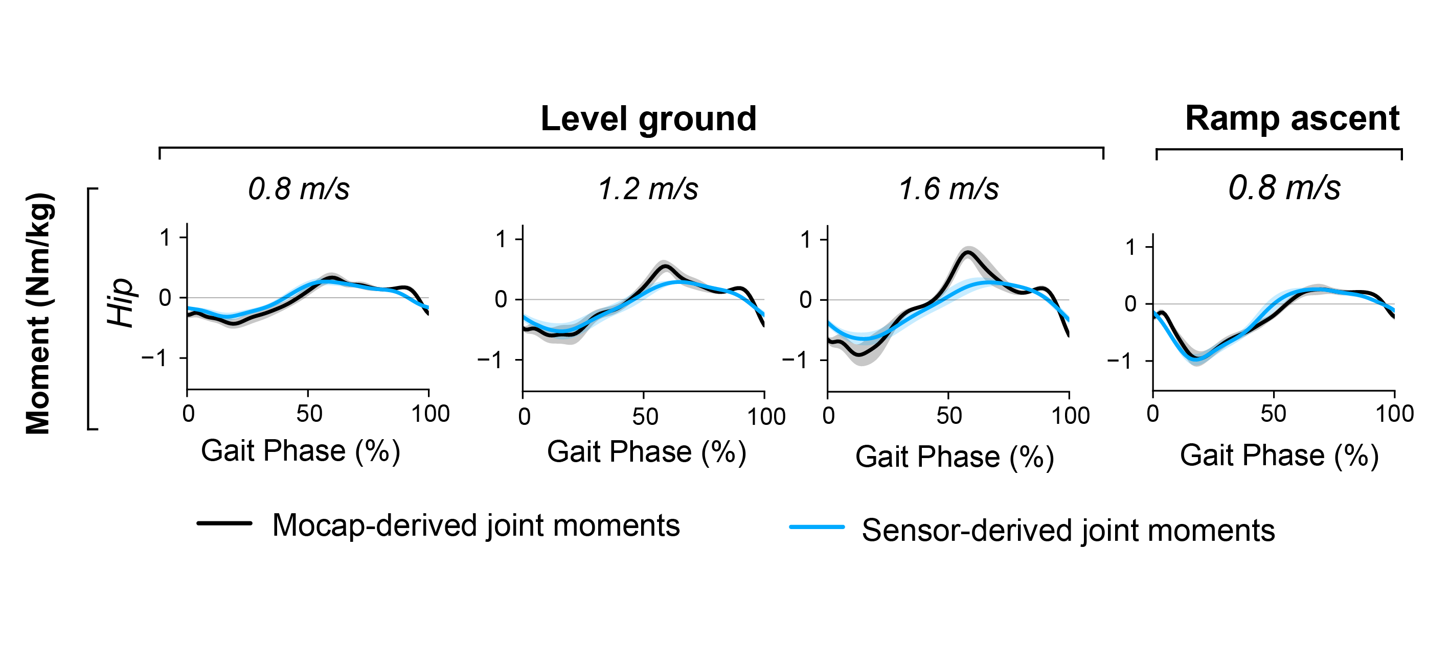
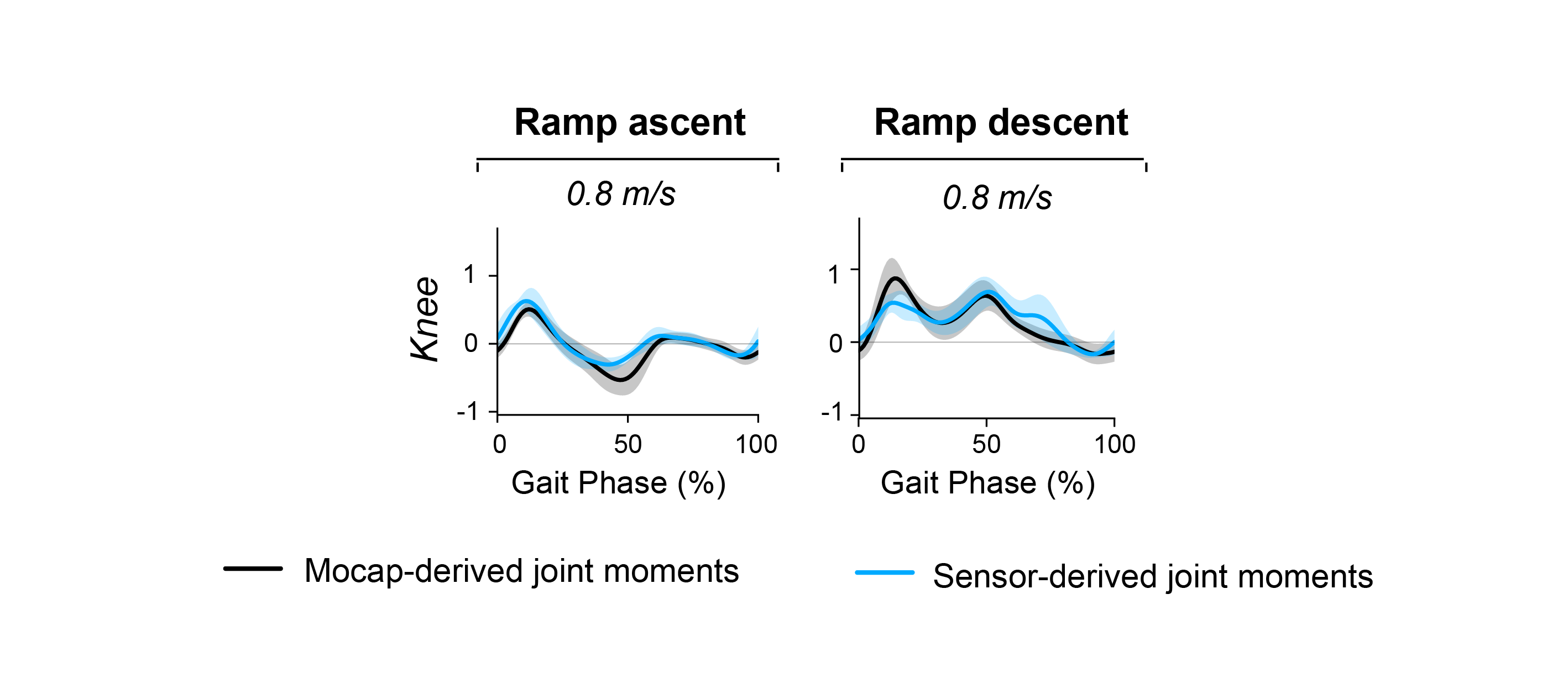
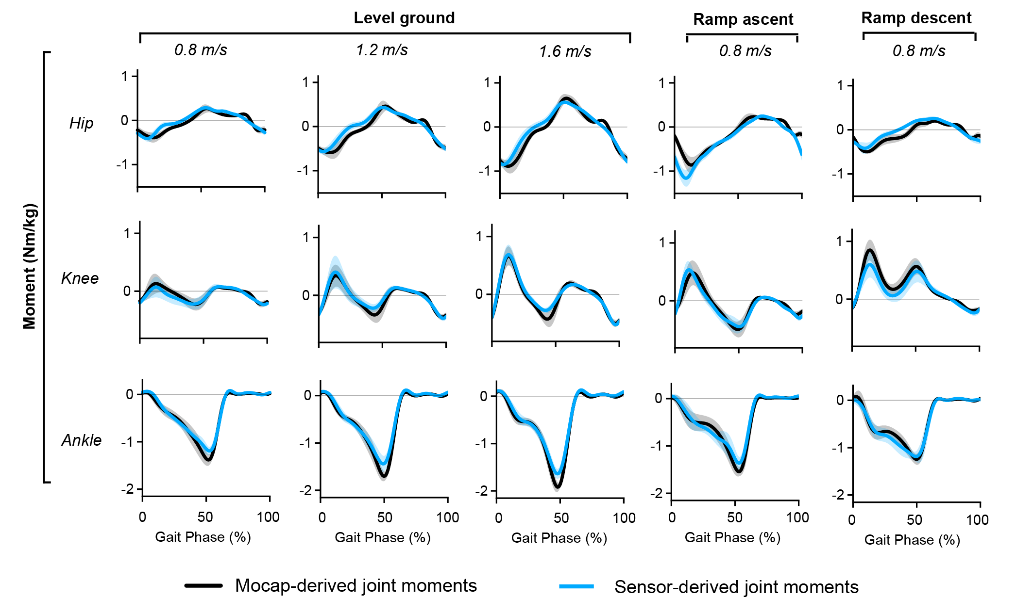
Objective: Data-driven models that estimate physiological states, particularly biological joint moments, are widely used in exoskeleton control. However, these estimators are often coupled to device-specific sensor configurations, limiting controller transfer and the use of open-source biomechanics datasets. Methods: We proposed joint kinematics as an intermediate representation that decouples hardware-specific sensing from downstream biological joint moment estimation. A joint-moment estimator using joint angles and angular velocities was trained exclusively on open-source biomechanics data and evaluated using kinematics from a hip exoskeleton, knee exoskeleton, and inertial measurement unit (IMU) sensor suite during level-ground, ramp-ascent, and ramp-descent walking. Results: The estimator achieved a root mean square error (RMSE) of 0.17 Nm/kg and coefficient of determination (R²) of 0.79 using hip exoskeleton kinematics, 0.19 Nm/kg and 0.62 using knee exoskeleton kinematics, and 0.15 Nm/kg and 0.85 using kinematics derived from IMUs across bilateral hip, knee, and ankle joints. Conclusion: Joint kinematics enabled an estimator trained only on open-source data to operate across distinct wearable platforms. Significance: This framework may reduce target-device data collection and support transferable biological joint moment estimation for exoskeleton control and wearable biomechanics.
A short walk-through of the unified kinematic representation and how it transfers across the hip exoskeleton, knee exoskeleton, and IMU sensor suite.
Video summary of the manuscript.
Citation
@misc{hwang2026unifiedkinematicrepresentationenables,
  title={A Unified Kinematic Representation Enables Reusable Biological Joint Moment Estimation},
  author={Jinwoo Hwang and Ilseung Park and Changseob Song and Vu Phan and Eni Halilaj and Inseung Kang},
  year={2026},
  eprint={2610.09150},
  archivePrefix={arXiv},
  primaryClass={cs.RO},
  url={https://arxiv.org/abs/2610.09150}
}